
Sometimes we don’t fail for lack of resilience, but because of biological overload. Based on a comprehensive systematic review, this new framework by researchers Juan Pablo Morales, Fiorella Macchiavello, and Felipe Rojas-Thomas maps how evidence indicates socioeconomic disadvantage is associated not just with psychological distress, but also with differences in our brainwaves, our stress hormones, and even our gut bacteria.
We often think of mental health as a deeply personal, psychological experience. But what if the roots of psychological distress are just as physical as they are mental, and can be explicitly measured in our brain waves, our blood, and our gut?
While we intuitively know that poverty and socioeconomic disadvantage (SED) are linked to stress, a recent systematic review dives into the potential biological mechanisms behind these relationships. The authors propose a new framework called CP-MInD (Contextual & Physiological Markers for Individual Distress) to explain how evidence suggests social adversity may physically embed itself into our biology and relate to our socio-cognitive abilities.
But how do researchers actually measure the biological correlates of a zip code or a bank account? By combining structural neuroimaging, real-time brainwave tracking, and multi-system biomarkers, scientists are mapping the physical footprint of inequality.
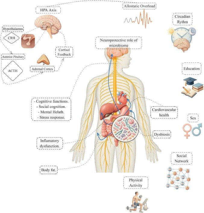
Figure 1. CP-MInD conceptual model linking contextual and physiological markers of individual distress. Adapted from Morales, J.P., Macchiavello, F., & Rojas-Thomas, F. (2026), Contextual & physiological markers for individual distress (CP-MInD). Brain health as a comprehensive framework for Mental-health equity, Neuroscience, 595, 154–170. Licensed under CC BY 4.0.
Tracking the “Wear and Tear”: Measuring Allostatic Load
To understand how context relates to the brain, researchers first look at how the body handles chronic stress through a metric called allostatic load (AL). AL essentially quantifies the biological “wear and tear” of surviving in a state of constant high alert.
Because chronic stress affects the whole body, measuring it requires a multi-system approach:
- Neuroendocrine Tracking: Researchers measure the Hypothalamic-Pituitary-Adrenal (HPA) axis by analyzing the “cortisol awakening response” (CAR) and diurnal cortisol slopes.
- Cardiovascular and Immune Panels: Alongside hormones, scientists track sympathetic-adreno-medullary activation (like blood pressure and catecholamines) and immunometabolic markers, such as C-reactive protein (CRP) and insulin resistance.
- Actigraphy: To capture how environmental stressors are associated with disrupted restorative processes, researchers use multi-night actigraphy (wearable sensors) to objectively track sleep fragmentation and circadian disruption.
Evidence indicates that when these biological systems are frequently activated in the presence of socioeconomic stressors, adaptive responses may become maladaptive, potentially leading to allostatic overload.
Mapping the Social Brain in Real-Time: Structural MRI and EEG
The CP-MInD framework emphasizes that socioeconomic disadvantage isn’t just associated with anxiety; evidence suggests it is fundamentally linked to differences in the neural circuits we use to navigate the social world. To observe this, researchers utilize two complementary neuroimaging tools:
- Structural MRI: Scans consistently reveal that chronic stress and systemic inflammation are associated with differences in prefrontal-limbic circuits. Individuals from lower-SES backgrounds, on average, show differences in gray matter volume and altered large-scale network organization in regions supporting cognitive control and emotion regulation.

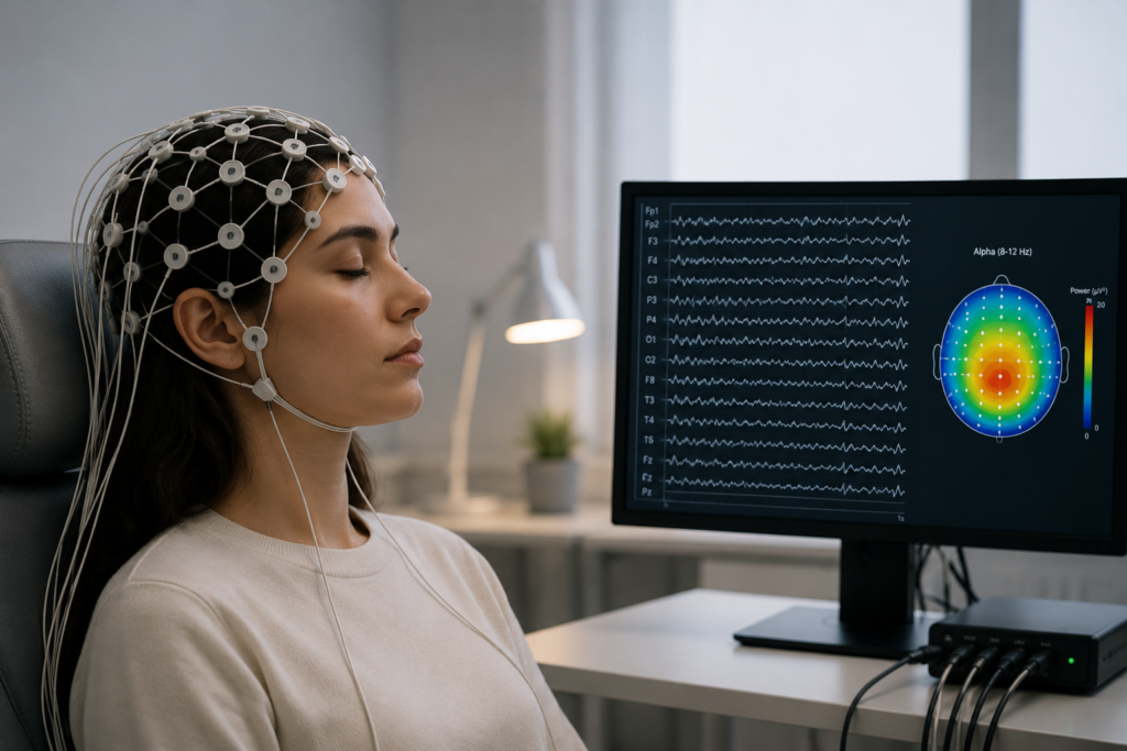
- Electroencephalography (EEG): While MRI shows the structure, EEG captures the brain in action. Researchers hook participants up to EEG caps while they perform social cognition tasks. By analyzing Event-Related Potentials (ERPs: specific spikes in brain activity like the P1, N1, N2, ERN, N400, and P3 components) scientists can track millisecond-level cognitive processing. The evidence shows that individuals from lower-SES backgrounds often display attenuated modulation of these components, which suggests an association with differences in early visual attention, conflict monitoring, and outcome evaluation.
Decoding the Gut-Brain Interface
Perhaps one of the most fascinating methodological frontiers in this research is the Microbiota-Gut-Brain Axis (MGBA). Disadvantaged environments frequently limit access to dense nutrition, often correlating with diets high in ultra-processed foods (UPFs).
By analyzing gut microbiota, scientists trace how microbially derived metabolites (like short-chain fatty acids) signal the brain via the vagus nerve, immune systems, and circulatory system. They measure markers of intestinal and blood-brain barrier permeability to explore how stress-related “dysbiosis” might allow inflammatory cytokines to affect neural resilience. The framework suggests this can be significantly associated with differences in social cognition and decision-making.
Moving Forward: The Power of Multimodal Research

If the biological correlates of socioeconomic disadvantage are this profound, understanding them requires more than just subjective self-reporting. The power of the CP-MInD framework is that it demands a multimodal methodology: pairing structural MRI and real-time EEG with multi-omics and physiological panels.
By tracking these objective markers, researchers are gathering evidence that navigating complex social landscapes under the weight of resource scarcity has a literal, measurable biological association. Ultimately, understanding these physiological pathways is essential for designing scalable, structural policies that don’t just address the symptoms of distress, but actively aim to mitigate the neurobiological links to inequality.
reference
Morales, J. P., Macchiavello, F., & Rojas-Thomas, F. (2026). Contextual & physiological markers for individual distress (CP-MInD): Brain health as a comprehensive framework for mental-health equity. Neuroscience, 595, 154–170. https://doi.org/10.1016/j.neuroscience.2025.12.034